
 Contatto
 Come acquistare
Consegna
Guida all'acquisto
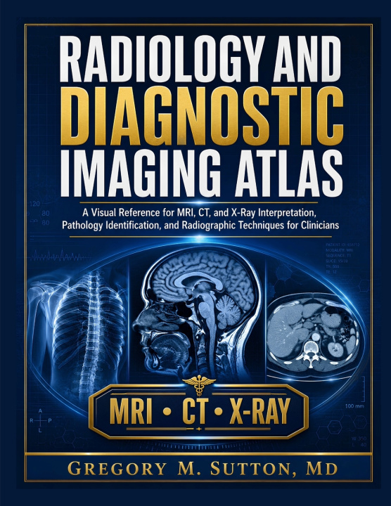
RADIOLOGY AND DIAGNOSTIC IMAGING ATLAS
A Visual Reference for MRI, CT, and X-Ray Interpretation, Pathology Identification, and Radiographic Techniques for Clinicians
 Inglese
 130 b
130 b
 Nuovi
Nuovi
Fino a 30 giorni per il reso
I clienti hanno acquistato anche




The Complete Visual Reference for MRI, CT, and X-Ray Interpretation-A Systematic Approach to Pathology Identification and Radiographic Techniques for Clinicians.
Diagnostic imaging is one of the most powerful tools in modern medicine, but interpreting images correctly requires more than pattern recognition-it demands a disciplined, repeatable approach to image review and a deep understanding of how each modality actually works. This comprehensive atlas is built to stand beside the clinician at the workstation, providing the physics fundamentals, systematic search patterns, and pathology identification frameworks needed to interpret X-ray, CT, and MRI studies with confidence.
The book begins with the foundations of image formation across modalities: X-ray physics and the five radiographic densities, CT acquisition and Hounsfield unit-based windowing, MRI sequence recognition including T1, T2, FLAIR, and diffusion-weighted imaging, and the complementary roles of ultrasound and nuclear medicine. You will learn the principles of radiographic positioning and technique standards, along with a systematic approach to image interpretation and structured reporting that reduces satisfaction of search error and other common interpretive pitfalls.
The organ-system chapters follow a consistent, easy-to-navigate structure: learning objectives, core teaching content, a systematic checklist you can apply at the workstation, common interpretive pitfalls, and a short clinical vignette illustrating the material in practice. Chest imaging covers systematic chest X-ray interpretation, CT evaluation of pulmonary nodules and infiltrates, cardiac and mediastinal imaging, and pleural pathology. Abdominal and genitourinary imaging addresses plain film and CT evaluation of the acute abdomen, solid organ pathology, GI tract obstruction and perforation, hepatobiliary and pancreatic imaging, and genitourinary and adrenal findings.
Musculoskeletal imaging covers fracture patterns and trauma radiography, arthritis differentiation by the four-feature framework, bone tumor characterization using age, location, margin, matrix, and periosteal reaction, spine imaging for degenerative, traumatic, and neoplastic findings, and pediatric musculoskeletal considerations. Neuroimaging and head and neck sections provide systematic approaches to stroke, hemorrhage, and trauma on non-contrast CT, intracranial masses and neurodegenerative patterns, head and neck space-based localization, and sinonasal and temporal bone imaging.
Breast and women's imaging covers mammography interpretation using the BI-RADS framework, obstetric and gynecologic ultrasound for early pregnancy viability, ectopic pregnancy, and adnexal mass characterization. Emergency and trauma imaging protocols, pediatric imaging considerations across modalities, contrast media safety and adverse reaction management, and the future of AI in diagnostic imaging complete the comprehensive coverage.
Whether you are a medical student, resident, practicing clinician, or allied health professional seeking a structured, visual reference for diagnostic imaging, this atlas is designed to build durable, transferable habits of image review. It is not a replacement for formal radiology training or institutional protocols-it is the systematic foundation that makes every subsequent read more confident and complete.


Informazioni sul libro
Inglese